The classic bunion, medically known as hallux abductovalgus or HAV, is a bump on the side of the great toe joint. This bump represents an actual deviation of the 1st metatarsal and often an overgrowth of bone on the metatarsal head. In addition, there is also deviation of the great toe toward the second toe. In severe cases, the great toe can either lie above or below the second toe. Shoes are often blamed for creating these problems. This, however, is inaccurate. It has been noted that primitive tribes where going barefoot is the norm will also develop bunions. Bunions develop from abnormal foot structure and mechanics (e.g. excessive pronation), which place an undue load on the 1st metatarsal. This leads to stretching of supporting soft tissue structures such as joint capsules and ligaments with the end result being gradual deviation of the 1st metatarsal. As the deformity increases, there is an abnormal pull of certain tendons, which leads to the drifting of the great toe toward the 2nd toe. At this stage, there is also adaptation of the joint itself that occurs.
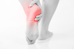
To understand the cause of the pain one must understand the anatomy of the foot and some basic mechanics in the function of the foot. A thick ligament, called the plantar fascia, is attached into the bottom of the heel and fans out into the ball of the foot, attaching into the base of the toes. The plantar fascia is made of dense, fibrous connective tissue that will stretch very little. It acts something like a shock absorber. As the foot impacts the ground with each step, it flattens out lengthening the foot. This action pulls on the plantar fascia, which stretches slightly. When the heel comes off the ground the tension on the ligament is released. Anything that causes the foot to flatten excessively will cause the plantar fascia to stretch greater than it is accustom to doing. One consequence of this is the development of small tears where the ligament attaches into the heel bone. When these small tears occur, a very small amount of bleeding occurs and the tension of the plantar fascia on the heel bone causes a spur on the bottom of the heel to form. Pain experienced in the bottom of the heel is not produced by the presence of the spur. The pain is due to excessive tension of the plantar fascia as it tears from its attachment into the heel bone. Heel spur formation is secondary to the excessive pull of the plantar fascia where it attaches to the heel bone. Many people have heel spurs at the attachment of the plantar fascia without having any symptoms or pain. There are some less common causes of heel pain but they are relatively uncommon.
There are several factors that cause the foot to flatten and excessively stretching the plantar fascia. The primary factor is the structure of a joint complex below the ankle joint, called the subtalar joint. The movement of this joint complex causes the arch of the foot to flatten and to heighten. Flattening of the arch of the foot is termed pronation and heightening of the arch is called supination. If there is excessive pronation of the foot during walking and standing, the plantar fascia is strained. Over time, this will cause a weakening of the ligament where it attaches into the heel bone. When a person is at rest and off of their feet, the plantar fascia attempts to mend itself. Then, with the first few steps the fascia re-tears causing pain. Generally after the first few steps, the pain diminishes. This is why the heel pain tends to be worse the first few steps in the morning or after rest. Another cause of heel pain is compression of the calcaneal nerve and may be diagnosed thru neuro sensory motor testing.
One other factor that contributes to the flattening of the arch of the foot is tightness of the calf muscles. The calf muscle attaches into the foot by the achilles tendon into the back of the heel. When the calf muscle is tight it limits the movement of the ankle joint. When ankle joint motion is limited by the tightness of the calf muscle it forces the subtalar joint to pronate excessively. Excessive subtalar joint pronation can cause several different problems to occur in the foot. In this instance, it results in excessive tension of the plantar fascia. Tightness of the calf muscles can be a result of several different factors. Exercise, such as walking or jogging will cause the calf muscle to tighten. Inactivity or prolonged rest will also cause the calf muscle to tighten. Women who wear high heels and men who wear western style cowboy boots will, over time, develop tightness in the calf muscles.
The next phase of treatment might consist of continued calf muscle stretching exercises, cortisone injections and orthopedic taping of the foot to support the arch. If this treatment fails, or if there is reoccurrence of the heel pain, then functional foot orthotics might be considered. A functional orthotic is a device that is prescribed and fitted by your foot doctor, which fits in normal shoes like an arch support. Unlike an arch support, however the orthotic corrects abnormal pronation of the subtalar joint. Thus orthotics address the cause of the heel pain – abnormal pronation of the foot.
The doctors at the Foot and Ankle Center are excited to also offer a new treatment, Extracorporeal Shock Wave Therapy, for chronic plantar fasciitis “heel pain”. Extracorporeal” means “outside the body”. Shock waves are created by very strong acoustic (sound) energy. Your ESW treatment will be performed with a device called the OssaTron. The OssaTron is a shock wave generator very similar to the shock wave devices used to treat kidney stones without surgery. The shock waves are created by a spark plus that is enclosed in a soft plastic dome filled with water. During ESW treatment, this dome is placed close against the heel so that the shock waves pass through the dome to the heel. ESW treatment has recently been found to be effective for treating chronic proximal plantar fasciitis.
Surgery to correct heel pain is generally only recommended if orthotics or the OssaTron treatment have not been successful. A new endoscopic treatment for heel pain is now available from Foot and Ankle Center of North Houston. This new method uses an endoscope which is a small camera instrument that allows the surgeon to see “anatomy” inside the body. By using a very small incision, less than ½ inch, this new procedure releases the extreme tension on the plantar fascia which is the cause of the pain in the majority of cases. All of this is viewed on the television monitor by the surgeon. The procedure itself usually takes less than 10 minutes using a local anesthetic. A sterile dressing is worn for approximately 3 days and then the patient is usually allowed to return to regular shoe wear. Minimal loss of work is incurred.
There are some exceptions to this course of treatment and it is up to you and our office to determine the most appropriate course of treatment. Following surgical treatment to correct heel pain the patient will generally have to continue the use of orthotics. The surgery does not correct the cause of the heel pain. The surgery will illuminate the pain but the process that caused the pain will continue without the use of orthotics. If orthotics have been prescribed prior to surgery they generally do not have to be remade.